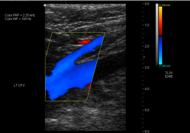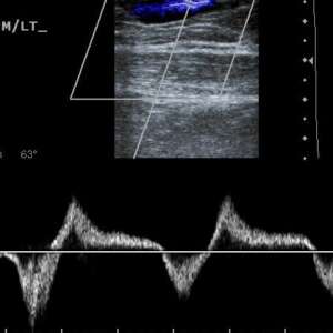
ULTRASOUND MONITORING DURING EVLT AND VNUS CLOSURE© OF THE GSV AND SSV Their position should be measured as distance (cm) from the floor in the extended limb.1820 The venous reflux examination also includes the mapping of exit and reentry perforating veins (PV).19 PV reflux is detected as outward flow duration greater than 350 ms on the release phase of flow augmentation maneuver (distal compression has higher sensitivity in detecting PVs reflux).1 Perforating veins should be accurately located in their different locations in the leg. Intersaphenous veins should also be identified and the variability in SSV termination carefully recorded. The diameters of the popliteal vein and the Small Saphenous vein (SSV) are recorded, as well as diameters of the SSV along its course in the leg. The Great Saphenous vein is then scanned in the leg and the thigh so that tributaries to the GSV should be noted (see Figure 23.1). They are easily confused with the GSV, especially during continuous longitudinal scanning when the saphenous vein appears to leave the saphe-nous compartment.18 Radiofrequency ablation commonly is applied to treat veins from 2-12 mm in diameter.16 The supragenicular, infragenicular, or immediate subgenicular Great Saphenous vein is often the access point for its laser or radiofrequency ablation.16,17 Therefore the depth of the GSV in these regions is additional data to be recorded.Īccessory veins by definition run parallel to the GSV in the thigh (see Figure 23.1).18 It is imperative to map their course accurately and to note their eventual communication with GSV (see Figure 23.1). The diameter of the saphenofemoral junction and femoral vein are recorded for use in judgment for radio frequency VNUS closure© and endovenous laser EVLT treatments.1416 Important information also is offered by the diameters of the GSV at mid thigh and distal thigh.

Patency usually is assessed by compression of the vein with the transducer.8 Reflux is detected by flow augmentation maneuvers such as distal compression and release of the thigh and calf or the Valsalva maneuver for only the saphenofemoral junction.8 Automated rapid inflation/deflation cuffs are cumbersome but may be used for this purpose and offer the advantage of a standardized stimulus.1012 Reflux greater than 500 ms is considered pathologic.9,13 Transverse and longitudinal scans combined with continuous scanning are performed in order to provide a clear mapping of the venous system. Measurements from the medial malleolus are not as precise. Their location measured in centimeters from the floor provides a therapeutic guide. Duplicated segments, sites of tributary confluence, and large perforating veins and their deep venous connections are identified. The veins are scanned by moving the probe vertically up and down along their course.

Sensitivity and specificity in detecting reflux are increased in examinations performed with the patient standing rather than when the patient is supine.8,9 Supine examinations for reflux are unacceptable. The ultrasound examination is conducted with the patient standing.9 This position has been found to dilate leg veins maximally and challenges vein valves. A clear graphic notation (mapping) of significant vein diameters, anomalous anatomy, superficial venous aneurysms, perforating veins, presence and extent of reflux should always be recorded during the examination (see Figure 23.1).2,5 From receiver operating characteristic (ROC) curve, optimal cut-off points of 27.8, 47.8, and 36.2 cm/s for the PRV at the SFJ ( p <0.01), GSV ( p <0.01), and SPJ ( p <0.01) discriminated between the two groups.Conclusion: PRV and MRV improved discrimination between early and advanced SVI compared to reflux duration.A detailed US duplex study of the normal and pathologic venous anatomy (reflux) is essential. MRV, PRV and TRV were greater in group II at the SFJ, SPJ and in GSV ( p <0.01 for all), although the duration of SPJ reflux was non-discriminatory ( p = 0.78). The vein diameter, reflux duration(s), mean reflux velocity (MRV cm/s), peak reflux velocity (PRV cm/s), and total reflux volume (TRV ml/s) were determined at the sapheno-femoral junction (SFJ), great saphenous vein (GSV) and sapheno-popliteal junction (SPJ).Results: Age and the proportion of males were greater in group II. Objectives: This study we compare the duplex-derived parameters of reflux in patients with early and advanced superficial venous insufficiency (SVI) to identify parameters reflecting this.Methods: Two thousand and one hundred sixty limbs with primary reflux, categorized according to the CEAP (clinical, etiologic, anatomic and pathophysiologic) classification, and the patients were divided into two groups (group I group II ) were studied.


 0 kommentar(er)
